Figure 8.

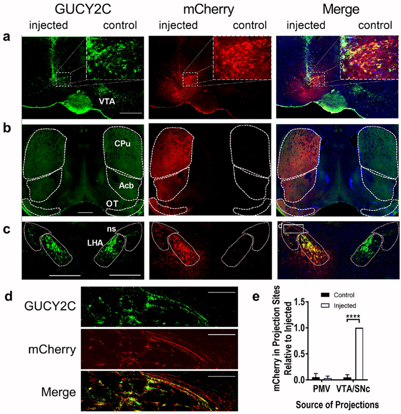
Selective anterograde tracing localized GUCY2C protein expression in VTA/SNc axons projecting to distant canonical regions, (a) Unilateral injection of AAV2-mCherry into the VTA produced mCherry expression in GUCY2C(+) neurons, (b-c) mCherry was co-expressed with GUCY2C in canonical ipsilateral projection sites of the VTA including the (b) CPu, Acb, and OT and (c) LHA and ns. (d) GUCY2C(+) axons from the VTA traveling in the ns express mCherry. (e) mCherry was quantitatively expressed in ipsilateral GUCY2C(+) projection sites of the VTA/SNc, but not in those of the PMV. Data in (e) represents mCherry expression normalized to maximum mCherry expression in each image. Data were collected from 2 mice with injection of mCherry into the VTA/SNc that spared the PMV. CPu: Caudoputamen; Acb: nucleus Accumbens; OT: Olfactory Tubercle; ns: nigrostriatal tract; LHA: Lateral Hypothalamic Area. Scale bars in (a-c): 200 μm, (d) 100 μm. ****p<0.0001.
