Figure 4.
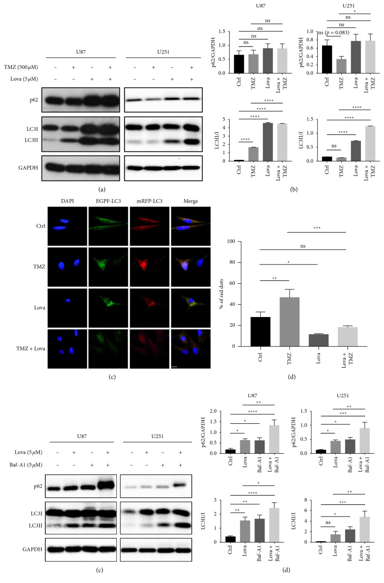
Lovastatin triggered autophagy but blocked autophagic flux in GBM cells. (a) U87 and U251 cells were treated with TMZ (500 μM), lovastatin (5 μM), or combination for 72 hrs, and autophagic markers (LC3I/II and p62) were detected by western blot. (b) Quantification of western blot results in (a); N = 3. (c) U87 cells expressing EGFP-mRFP-LC3 were treated with TMZ (500 μM), lovastatin (5 μM), or combination for 72 hrs. Fluorescence images were taken by using a confocal microscope (scale bar = 20 μm). The average number of red and yellow puncta per cell was calculated from three random-picked view fields. (d) Quantification of results in (c); N = 3. (e) U87 and U251 cells were treated with lovastatin (5 μM), bafilomycin-A1 (5 μM), or combination for 72 hrs, and autophagic markers were detected by western blot. (f) Quantification of western blot results in (e); N = 3. ∗p < 0.05; ∗∗p < 0.01; ∗∗∗p < 0.001; ∗∗∗∗p < 0.0001; ns = no significance.

