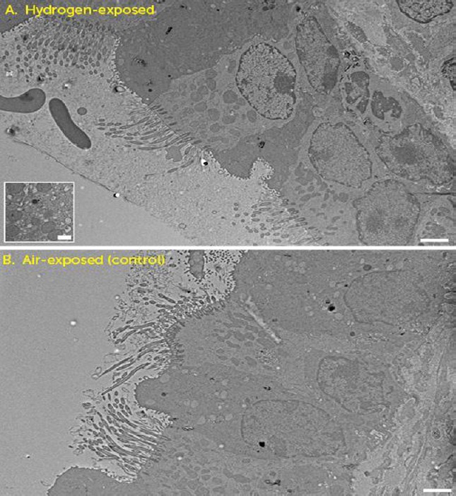
Figure 4.

Electron microscopic analysis of the hydrogen- (A) and air-exposed (B) revealed that the micro- and macro-structures of airway epithelial cells were normal.
Note: (A, inset) Two animals exposed to hydrogen gas exhibited a prominence of secretory vesicles in respiratory epithelium, though nuclear, mitochondrial and ciliary structures remained intact. Scale bars: 2 μm.
