Figure 3.

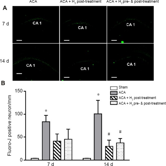
Effect of hydrogen (H2) treatment on the neuronal survival in the hippocampal CA1 region of rats with asphyxia induced-cardiac arrest (ACA) at 7 and 14 days (d) post-resusciation.
Note: (A) In rats subjected to ACA, there were signifcantly greater number of Fluoro-Jade staining positive neurons, suggesting neuronal degeneration. H2 treatment improved neuron survival, which was significant at 14 d post-resuscitation. (B) Quantitative result of Fluoro-Jade staining positive neurons. Data were presented as the mean ± SEM (n = 6/group). *P < 0.05, vs. sham group; #P < 0.05, vs. ACA group (one-way analysis of variance followed by Student-Newman-Keuls post hoc test).
