
Figure 1.
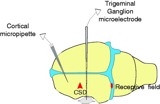
Experimental setup. Diagram showing position of: Cortical micropipette for electrocorticogram recording; trigeminal ganglion microelectrode, which was advanced through the contralateral (right) cortex with a medial angle to reach the left trigeminal ganglion, for unit recording; and a representative mechanical receptive field on the transverse sinus.