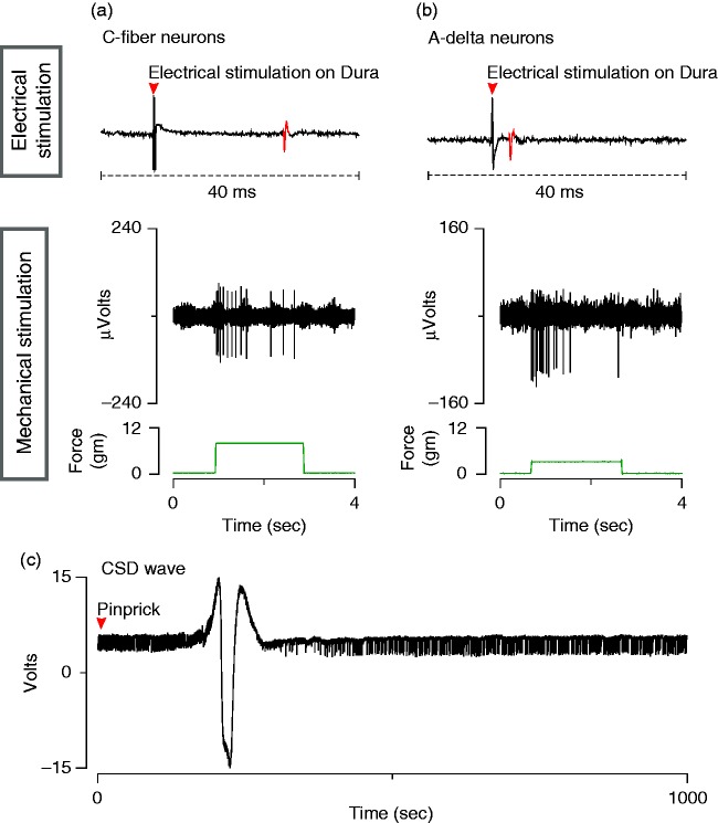
Figure 2.
Electrophysiological characterization of meningeal nociceptors and CSD. (a), (b). Electrophysiological recordings of action potentials in C-fiber (a) and Aδ-fiber (b) meningeal nociceptors evoked by electrical and mechanical stimulation of their receptive fields on the transverse sinus. (c) Electrocorticogram showing CSD wave induced by pinprick in the cortex.

