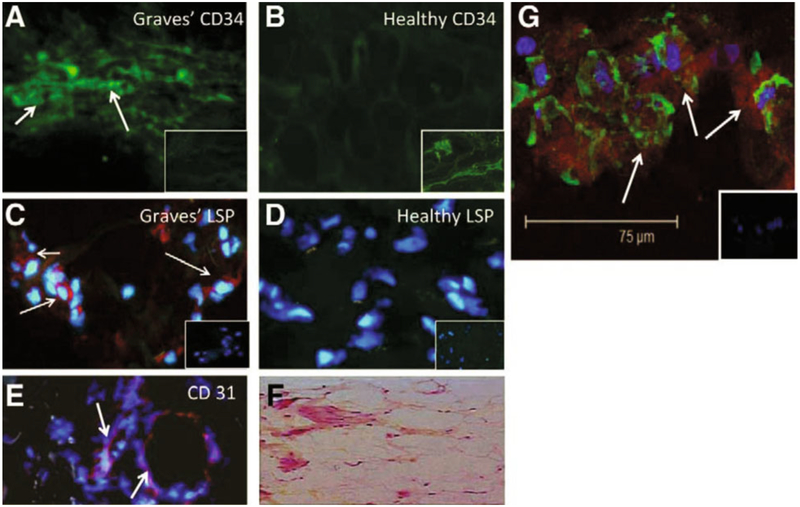
Fig. 1 -.
CD34+ LSP-1+ TSHR+ fibrocytes can be identified in the orbital tissue of patients with thyroid-associated ophthalmopathy (TAO) but are absent in tissues from healthy donors. A) CD34 expression (arrows, green FITC) in TAO-derived tissue (inset, negative control staining). B) Absent CD34 expression in healthy tissue (inset, positive staining control). C) LSP-1 expression in TAO-derived tissue [red, arrows, nuclei counterstained with DAPI (blue)] (inset negative control). D) Absence of LSP-1 expression in healthy tissue (inset negative control). E) CD31 expression in disease-derived tissue is limited to vascular endothelium (red, arrows). F) H and E stained consecutive thin-sections of the same orbital tissue (40 x). G) Fibrocytes present in orbital tissue from patients with TAO co-express CD34 and TSH receptor (TSHR). Thin sectioned tissue from a donor with TAO was stained according to procedures described in “Methods” with anti-CD34 (green) and anti-TSHR (red) antibodies. Nuclei were counterstained with DAPI (blue). Thin sections were then subjected to confocal microscopy. Inset contains a negative staining control. [Reprinted with permission from (32), copyright 2010, The Endocrine Society].

