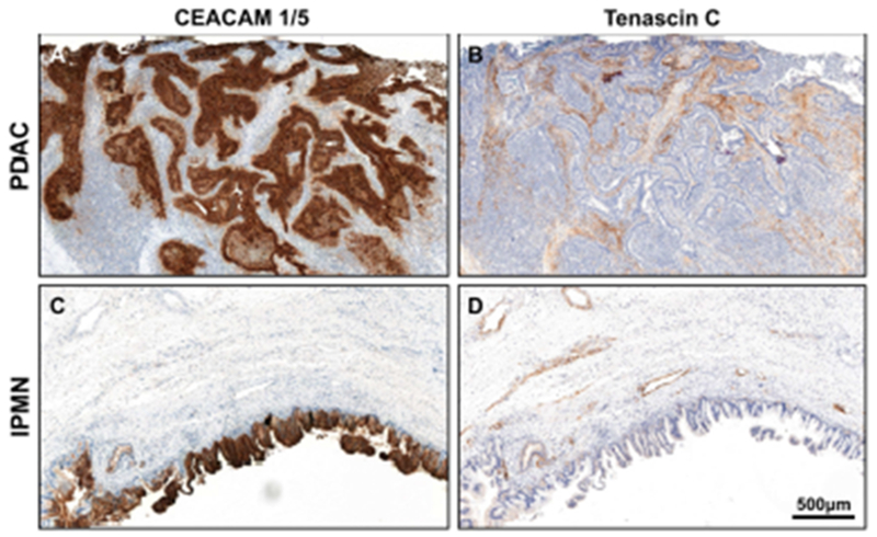
Fig. 2.

A representative PDAC with CEACAM 1/5 staining that is strong and diffuse in the tumor epithelial cells (A), whereas tenascin C staining that is strong and diffuse in the stroma (B). A representative IPMN with CEACAM 1/5 staining that is strong and diffuse in the tumor epithelial cells (C), whereas tenascin C staining that is moderate and focal in the stroma (D).
