Figure 5.
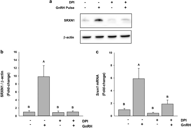
DPI attenuated GnRH pulse–mediated SRXN1 and PRDX3 induction. LβT2 cells (n = 1 × 10⁷) were cultured on Cytodex microcarriers for 24 h in DMEM/10% FBS with antibiotics. The cells were changed into serum-free medium and incubated for an additional 16 h. Cells were pretreated with vehicle or 5 μM DPI for 30 min and then pulsed for 2 min at 58-min intervals with vehicle or 10 nM GnRH for 4 h. After pulse stimulation, total RNA and protein were harvested. Protein was analyzed by western blot to detect (a) SRXN1 protein levels and (b) PRDX3 oxidation. (c) Intensity values for SRXN1 were measured by quantitative chemiluminescence and charted. (d) Isolated RNA was evaluated by qPCR for Srxn1 levels; charts represent mean values ± SEM. Different letters indicate a significant difference between groups (P < 0.05) by two-way ANOVA and post hoc testing with the Tukey multiple-comparison test.

