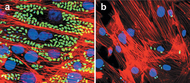
Figure 3. GKV25 strongly inhibit T. cruzi myoblast infection at 3.5 nM.
Cardiomyocyte monolayers were infected with GFP-expressing trypomastigotes of the Tulahuen for 24 h and then treated with different concentrations of GKV25 dissolved in DMSO. Controls were infected cells exposed to the same concentrations of DMSO. Parasite multiplication within monolayers was evaluated by fluorescence confocal microscopic observations of T. cruzi parasites inside cardiomyocytes 72 h after infection as described [20]. T. cruzi amastigotes are green, cardiomyocyte nuclei are blue, and cardiomyocyte actin filaments are red. Panel a shows control infected cells treated with DMSO whereas panel b shows the effect of GKV25 on infected myoblasts at 3 nM.

