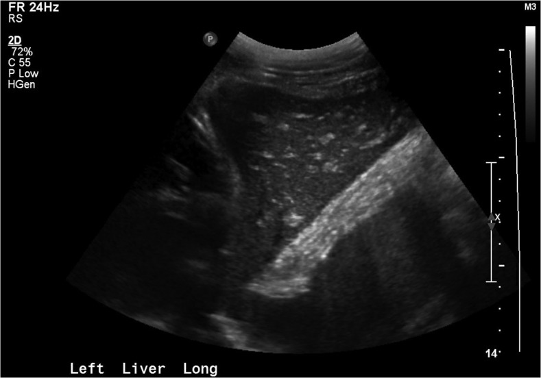
Figure 2.
Case 2: Right upper quadrant ultrasound with “starry sky” appearance, consistent with acute liver injury (echogenic portal triads and venous walls with hypo-echoic, edematous liver parenchyma). This pattern has not only been most commonly reported in viral hepatitis but has also been observed in heart failure, and other infectious, inflammatory, and neoplastic conditions of the liver.20

