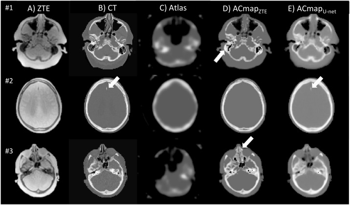
Fig 1. Axial sections of ZTE MR image (A), CT that served as reference (B), AC map generated from Atlas (C), AC map generated from segmented-ZTE (D), U-net method (E) in three different patients.
With ACmap ZTE (patient #1, ZTE-based AC yields to a global overestimation of attenuation in the ethmoids or mastoids cells as shown in D (arrow). In patient #3, ZTE-based AC yields to a global overestimation of attenuation in the ethmoids or mastoids cells as shown in D (arrows). With ACmapU-net, (E) mastoids cells were often filled with blurry structures. The calcification of a small meningioma of the falx cerebri found in patient #2 is partly restored with the ACmapU-net (arrow) opposite to ZTE- AC.

