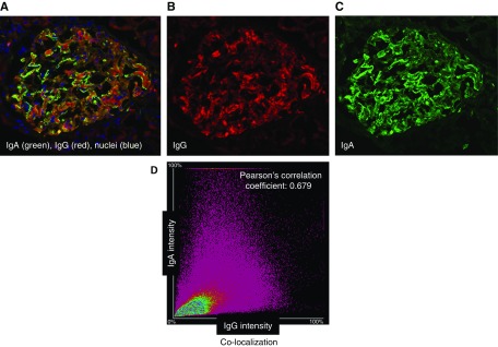
Figure 3.
Confocal microscopic analyses of glomerular Ig deposits of a patient with IgAN with IgG by routine immunofluorescence microscopy show colocalization of IgG and IgA. A remnant frozen kidney-biopsy specimen from a patient with IgAN with IgG staining by routine immunofluorescence microscopy was stained with antibodies against IgA (Cy2, green) and IgG (Alexa 555, red), and a DNA stain (DAPI, blue). (A–C) Single-layer confocal microscopy images with three combined colors (A), red (B), and green (C). (D) Image analysis of entire glomerular area indicated colocalization of individual components, IgA and IgG in glomerular deposits. For this sample, Pearson correlation coefficient (r) is 0.68. Pearson correlation coefficient (r) with value of 1.0 would indicate 100% colocalization.

