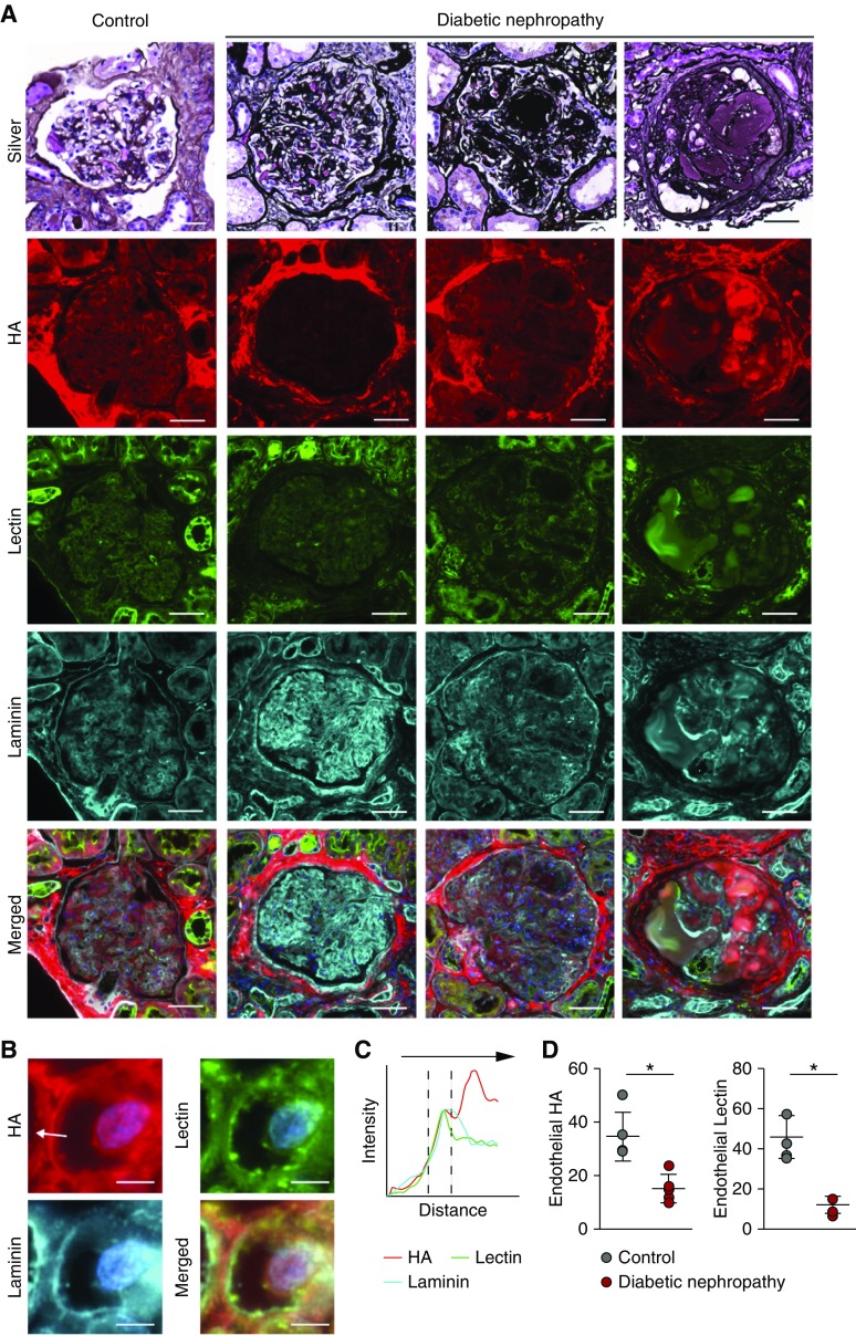
Figure 2.
Early-stage human diabetic nephropathy shows loss of glomerular HA. (A) Representative methenamine silver–periodic acid–Schiff–stained glomeruli of a control glomerulus (left panel) and three consecutive stages of DN in human kidney biopsy specimens. Further, fluorescent images of glomerular tissue sections stained for HA (red, second row), Lycopersicon esculentum lectin (green, third row), and laminin (cyan, fourth row), and merged images. (B) Insets show examples of measurement lines (scale bar, 5 µm). (C) Fluorescence intensity plots demonstrate endothelial localization of HA in relation to the subendothelial basement membrane position (cyan, laminin). (D) Quantification of total endothelial HA (left) or lectin (right) presence (n=5 per group).

