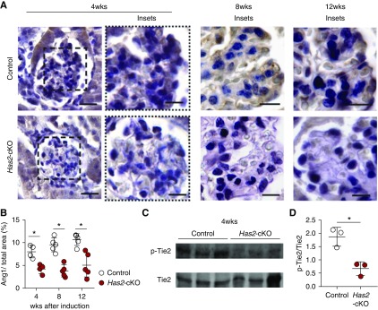
Figure 5.
Loss of HA leads to reduction of Ang1/Tie2 signaling in vivo. (A) Representative images of glomerular angiopoietin 1 (Ang1) presence at 4 weeks (left panels) after tamoxifen induction in control (upper panels) and has2-cKO (lower panels) mice (scale bar, 60 µm) with detailed glomerular tufts at 4, 8, and 12 weeks (scale bar, 30 µm). (B) Quantification of glomerular tuft Ang1 (n=5 per group). (C) Western blot samples from kidneys of Has2-cKO and control mice at 4 weeks stained for p-Tie2 and Tie2. (D) Quantification of p-Tie2(Y992) over Tie2 expression. Values are given as mean±SEM or mean±SD and difference was assessed by nonpaired two-tailed t test; *P<0.05.

