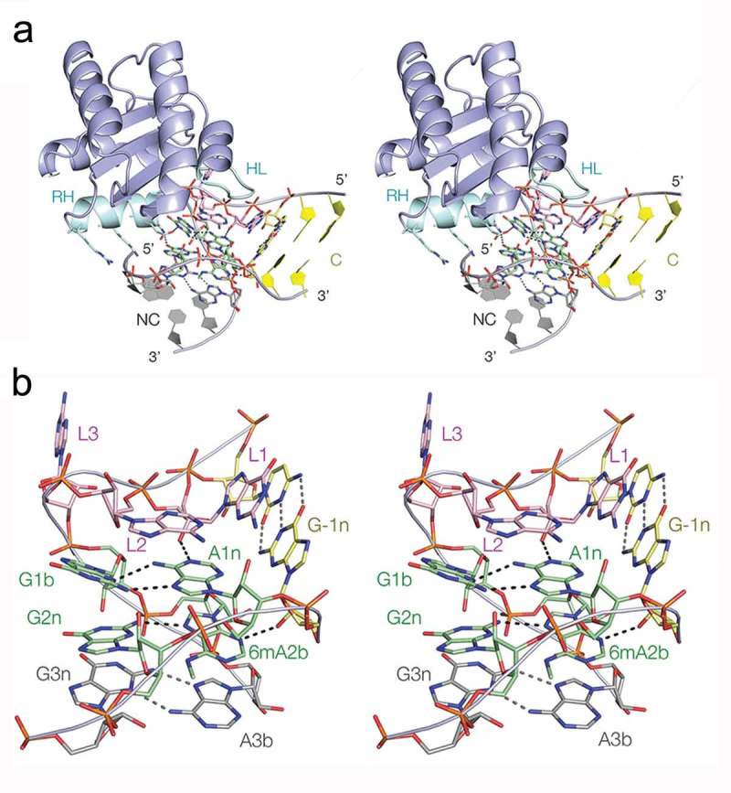
Figure 5.

The crystal structure of Kt-7 with N6-methyladenine at the 2b position bound to AfL7Ae. Parallel-eye stereoscopic views are shown.
(a). Overall view of the structure seen from the side of the non-bulged strand. The recognition helix (RH) and hydrophobic loop (HL) of the protein are highlighted in cyan. Side chains of the recognition helix that interact with the RNA are shown in stick form. (b). The structure of the core of the k-turn in the complex. The RNA adopts a standard N3-conformation k-turn structure.
