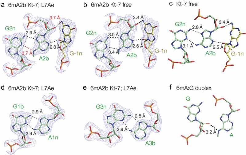
Figure 6.

A2b:G2n base pairs found in different structures and positions. For those taken from the current study, the 2Fo-Fc electron density map contoured at 1.2 σ is shown.
(a). The 2b:2n.-1n interaction in the structure of the complex of A2n N6-methyladenine Kt-7 with AfL7Ae. Potential hydrogen bonds are shown as broken lines, with distances > 3.3 Å drawn red. (b). The 2b:2n.-1n interaction in the structure of protein-free A2n N6-methyladenine Kt-7. (c). The 2b:2n.-1n interaction in the structure of protein-free unmodified Kt-7 (PDB 4CS1) [39]. (d). The 1b:1n interaction in the structure of the complex of A2n N6-methyladenine Kt-7 with AfL7Ae. (e). The 3b:3n interaction in the structure of the complex of A2n N6-methyladenine Kt-7 with AfL7Ae. (f). The G:A interaction in an RNA duplex containing tandem G:A, A:G base pairs flanked by G:U base pairs (PDB 5LR3) [15]. Note that in this structure there is no hydrogen bonding between the two nucleobases.
