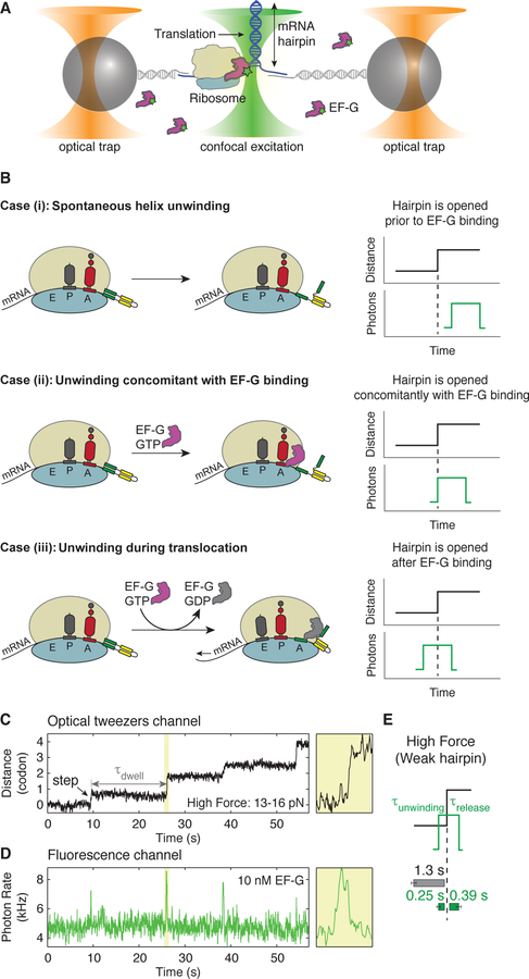
Figure 1. Ribosome opens an mRNA hairpin after EF-G binding.
(A) Experimental setup for simultaneous measurement of hairpin opening and EF-G binding using a time-shared optical trap with single-molecule confocal imaging, ‘fleezers’. Time resolution of the optical tweezers channel is 7.5 ms and that of fluorescence channel is 10 ms. Assembly of ribosome-stalled complexes is outlined in Figure S1.
(B) Possible scenarios of when hairpin unwinding occurs with respect to EF-G binding.
Case (i): the hairpin is spontaneously opened prior to EF-G binding due to the destabilization energy contributed by the ribosome (Qu et al., 2011).
Case (ii): the hairpin is opened concomitant with EF-G binding. Here, either EF-G binding itself induces a conformational change (for example, the forward or reverse movement of the 30S head) that results in the opening of the hairpin, or the 30S head rotates back and forth as a Brownian ratchet, and only the binding of EF-G effectively rectifies the position of the 30S head leading to hairpin opening.
Case (iii): the hairpin is opened a variable amount of time after EF-G binding. Opening could result from events such as EF-G•GTP tight binding, GTP hydrolysis, Pi release, EF-G•GDP release or internal conformational changes of the ribosome.
(C, D) An example of a single-ribosome ‘fleezers’ trajectory along an mRNA hairpin held under high external force of 13–16 pN with 10 nM Cy3-labeled-EF-G. (C) Optical tweezers channel, ribosome opens the hairpin in 1 codon steps (= 6 nts of hairpin opened) separated by dwells, τdwell. Data was recorded at 133 Hz and displayed at 13 Hz. Yellow box shows a zoomed-in event. (D) Fluorescence channel, each spike in fluorescence corresponds to the binding of an EF-G. Yellow box shows a zoomed-in event. Data is recorded at 100 Hz and displayed at 10 Hz. Additional zoomed-in events shown in Figure S2.
(E) Summary of average τdwell (grey) and average EF-G residence times before unwinding (τunwinding, green) and after unwinding (τrelease, green) for a weak hairpin held under external force of 13–16 pN (n = 55 events, 9 molecules). Error bars represent SEM.

