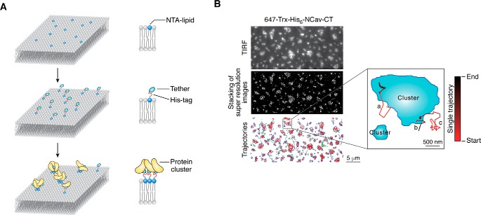
Figure 4.
Condensed phase formed on 2D supported lipid bilayer. A, schematic diagram of microdomain formation on 2D supported lipid bilayer. Membrane proteins homogeneously distribute on the supported lipid bilayer via tethering of the His-tag to Ni2+-NTA-decorated lipids. Protein clusters are observed on lipid bilayers after the addition of other components to drive phase separation. B, STORM analysis of membrane proteins, the cytoplasmic tail of NCav as an example, on supported lipid bilayer (adapted from Ref. 45). Image captured under TIRF microscopy mode first sketches the contours of the condensed phase, which turns out to perfectly overlap with the image reconstructed from STORM analysis. Trajectories of individual molecules are followed by single molecular tracking assay, both inside and outside the condensed phase. Direction of movement is marked by gradient color from black to red.

