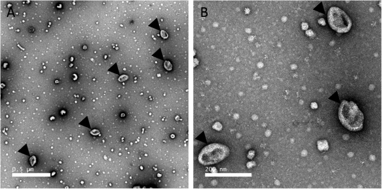
Figure 8.
Transmission EM of EV fractions purified from uterine fluid harvested at 7 h post-ovulation. A, low magnification view. B, higher magnification TEM micrograph of the EVs. EVs were negatively stained with 2% uranyl acetate (see “Experimental procedures”). Black arrowheads indicate extracellular EVs. Scale bars, A, 0.5 μm; B, 200 nm.

