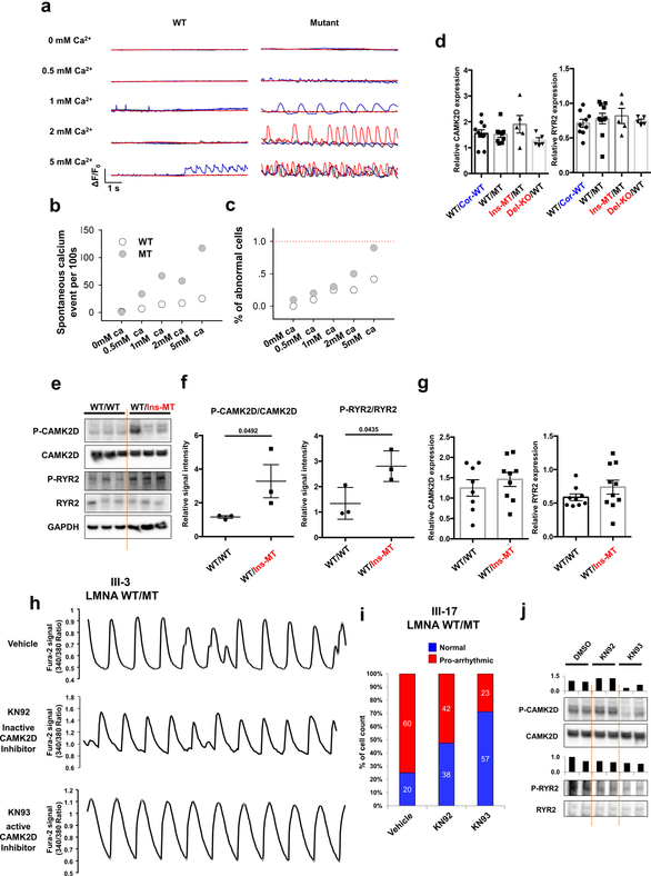
Extended Data Fig. 3. Abnormal calcium handling in LMNA mutant iPSC-CMs.
a, Confocal imaging of Fluo-4AM calcium events in control (III-3; WT/Cor-WT) and mutant (III-3; WT/MT) iPSC-CMs while being treated with increasing extracellular Ca2+ concentration. All representative traces were the recordings from 3 individual cells (presented as red, blue, and black). b, Spontaneous calcium event per 100s of control and mutant iPSC-CMs in each different extracellular Ca2+ concentration. c, Summary of the percentage of cells exhibiting spontaneous SR Ca2+ release events in control and mutant iPSC-CMs. d, Real-time analysis of CAMK2D and RYR2 expression in control and mutant iPSC-CMs. Data are expressed as mean ± s.e.m. e, f, Immunoblot analysis of phospho-RYR2, RYR2, phospho-CAMK2D, and CAMK2D protein levels in control and mutant iPSC-CMs. Data are expressed as mean ± s.e.m., and a two-tailed Student’s t-test was used to calculate P values. n=3. Numbers above the line show significant P values. g, Real-time PCR analysis of CAMK2D expression in control and mutant iPSC-CMs. Expression level of GAPDH was used as control. Data are expressed as mean ± s.e.m., and a two-tailed Student’s t-test was used to calculate P values. n=8. h, Representative Ca2+ transients of mutant iPSC-CMs (III-3; WT/MT) treated with 1 uM of KN92 or KN93 for 24 hr. i, Quantification of the percentage of cells exhibiting arrhythmic waveforms in mutant iPSC-CMs (III-17 and WT/MT) at basal level, as well as after the treatment with 1 uM of KN92 or KN93 for 24 hr. j, Immunoblot analysis of phospho-RYR2, RYR2, phospho-CAMK2D and CAMK2D protein levels with treatment of DMSO, KN92 or KN93 for 24 hr. The experiments in a were repeated twice independently with similar results. The Ca2+ transients in h were repeated as described in figure 2e independently with similar results. The Immunoblot data in e and j was repeated twice independently with similar results.

