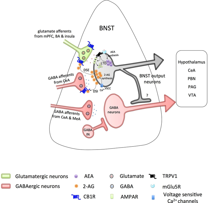
Figure 2. Endocannabinoid signaling in the bed nucleus of the stria terminalis (BNST).

Excitatory inputs (light green) from medial prefrontal cortex (mPFC), basal amygdala (BA) and insula, and inhibitory inputs (light red) from central nucleus of the amygdala (CeA) project onto BNST neurons and are regulated by the endocannabinoid system via presynaptic CB1 receptors. Inhibitory inputs from CeA and medial amygdala (MeA) projecting onto GABAergic BNST neurons (light red) are devoid of CB1 receptors. Activity at excitatory inputs evokes postsynaptic depolarization of BNST neurons, subsequent Ca2+ entry via voltage sensitive calcium channels (VSCC), initiates the production of 2-arachidonoyl glycerol (2-AG), activation of presynaptic CB1 receptors at both excitatory and inhibitory inputs, in turn, resulting in short-term suppression of glutamate (depolarization-induced suppression of excitation, DSE) and GABA (depolarization-induced suppression of inhibition, DSI) release. Activation of mGluR5 receptor initiates the production of anandamide (AEA), which further activates the postsynaptic TRPV1 channels in an autocrine manner. TRPV1 triggers internalization of postsynaptic AMPA receptors and induce long-term depression (LTD). BNST neurons send dense projections to the lateral hypothalamus, CeA, parabrachial nucleus (PBN), periaqueductal gray (PAG), and ventral tegmental area (VTA).
