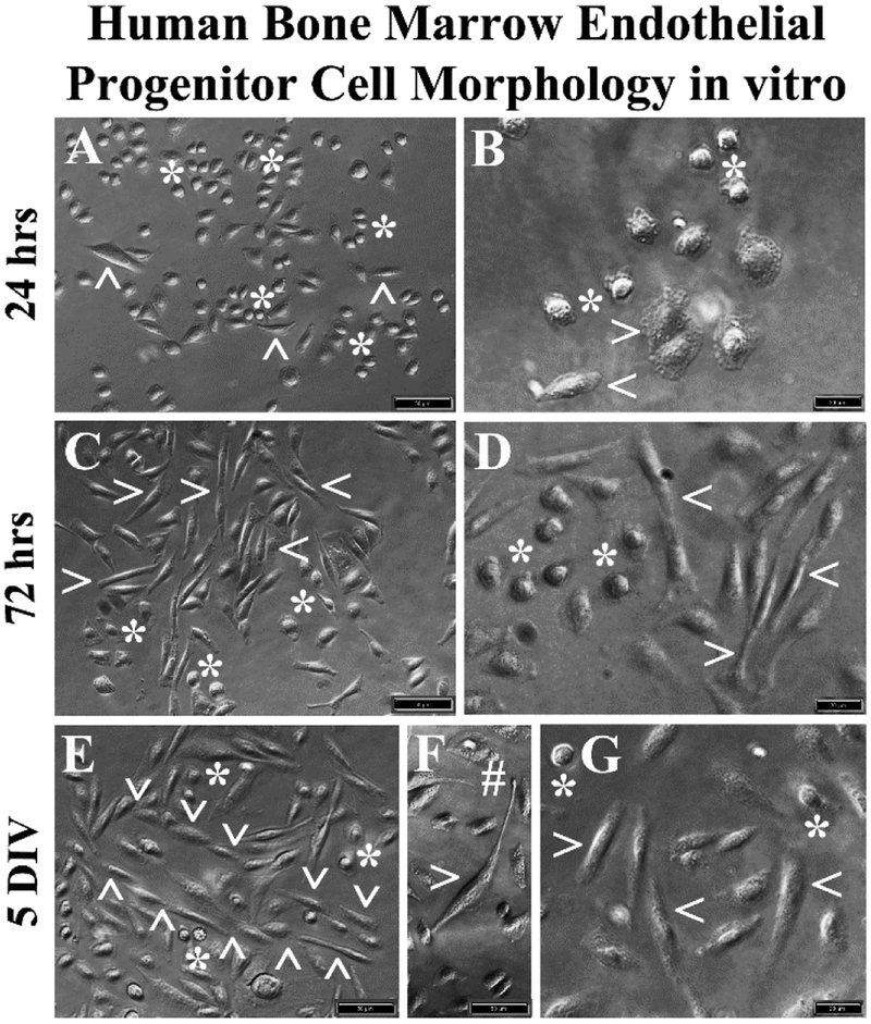
Figure 2. Phase-contrast images of hBMEPCs in vitro at different time points.
(A, B) The hBMECs showed mainly morphologically rounded cells (asterisks) and a few elongated cells (arrowheads) were observed at 24 hrs of culture in basal media. (C, D) More elongated cells (arrowheads) were detected in cultures at 72 hrs. Numerous rounded cells (asterisks) were noticed. (E) At 5 DIV, a tubular-like vessel formation (arrowheads) composed of elongated cells was determined. (F) Elongated cells also displayed long processes with tip development (hashtag). (G) A few rounded cells (arrowheads) were still observed at 5 DIV. Scale bar in A, C, E, F is 50 μm; in B, D, G is 20 μm.

