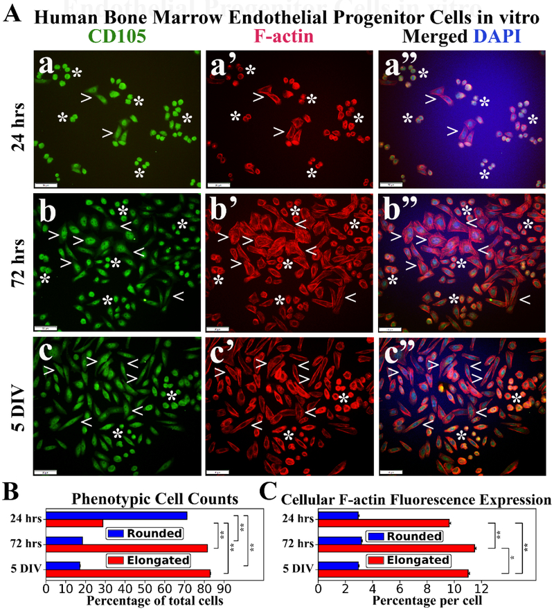
Figure 3. Immunocytochemical analyses of hBMEPCs in vitro at different time points for CD105 and F-actin expressions.
(A) CD105 immunoexpression was detected in all cells at each time point (green, a, b, c). During post-culture, F-actin immunoexpression (red) displayed diffusely within cytosol of rounded hBMEPCs (arrowheads) and elongated cells (arrowheads) exhibited organized filamentous strips in cell cytoplasm (a’, b’, c’). Merged images (a”, b”, c”) are shown with DAPI (blue). Scale bar in a-c” is 50 μm. (B) Rounded hBMEPC prevalence significantly decreased in conjunction with significantly increased occurrence of elongated hBMEPC from 24 hrs to 5 DIV. (C) Although F-actin immunoexpression levels remained stable over time, a significant increase of cytoskeletal filaments was determined in elongated cells from 24 hrs to 5 DIV. *p < 0.05, **p < 0.01.

