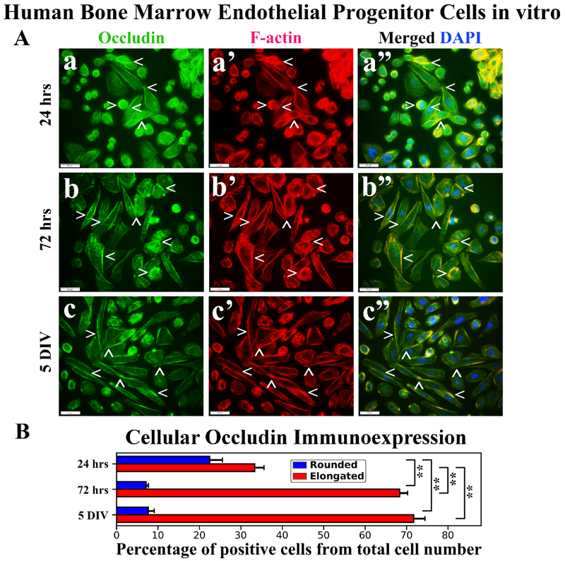
Figure 5. Immunocytochemical analysis of hBMEPCs in vitro at different time points for occludin and F-actin.
(A) Occludin immunoexpression in rounded hBMEPCs localized on cell membrane surfaces and elongated cells showed this protein immunoexpression as strips between adjacent cells (green, arrowheads, a, b, c). Occludin immunoexpression appeared to be distal from F-actin in numerous cells (red, arrowheads, a’, b’, c’). Merged images (a”, b”, c”) are shown with DAPI (blue). Scale bar in a-c” is 50 μm. (B) Percentage of rounded occludin immunopositive hBMEPCs substantially decreased from 24 hrs to 5 DIV. Significantly increased percentage of elongated hBMEPCs immunoexpressing occludin was determined from 24 hrs to 5 DIV. **p < 0.01.

