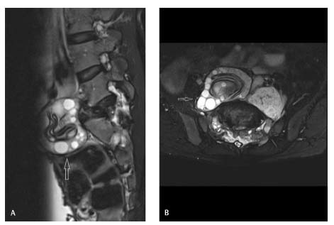
Fig. 1.

T2-weighted fat-suppressed sagittal ( A ) and axial (B) magnetic resonance images show an expansile lesion (arrows) with peripherally arranged hyperintense cysts and centrally clumped curvilinear hypointense membranes, causing collapse of S1 vertebra and extending into the prevertebral and paravertebral regions around L5 to S2 vertebrae.
