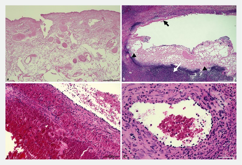
Fig. 2.

Histologic findings of resected specimen from mini-pigs with periductal injury. a RFA applied area reveals severe coagulated necrosis of bile duct without viable structure. b Damaged bile duct shows mucosal coagulative necrosis (black arrow), total lysis of ductal wall (arrow head) and periductal abscess (white arrow). c In some portal veins, neutrophilic phlebitis, endothelial degeneration, and marked intra- and peri-venous hemorrhage are present. d Adjacent hepatic artery shows prominent neutrophilic infiltration associated with necrosis and hemorrhage of the arterial wall.
