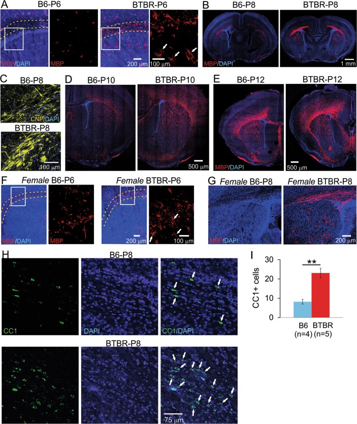
Fig. 2. Increased myelination during neonatal development in the frontal brain of both male and female BTBR mice.
a At P6, MBP signal was located in many cells in male BTBR frontal brain (right panels), including corpus callosum and dorsal striatum, and to a less extent, the cortex. Most of the positive cells had MBP signal in both cell bodies and numerous processes, with some already aligned in parallel tube-like structures presumably wrapping around axons. On the contrary, only a few MBP-positive cells could be identified in the same brain region in the B6 brain (left panels), with the signal mostly restricted to the cell bodies. Dotted lines outline corpus callosum. Boxed region is shown in higher magnification in the right panel next to it. (b) At P8, MBP signal in the BTBR frontal brain showed clear organization of myelin sheath in the corpus callosum and dorsal striatum, with dispersed positive cells in the cortex, while the same staining showed mostly scattered signal in the same brain regions in the B6 mice. Similar difference was observed in CNP immunofluorescence staining as well (c). d, e At P10 and P12, MBP signal continued to increase rapidly in both strains; however, it spread further into more superficial regions of the cortex and ventral striatum in the BTBR than the B6 brain. Similarly increased MBP signal in female BTBR brains during neonatal development was observed at P6 (f) and P8 (g). n = 2 to 4 for each strain at each age of each sex. White arrows in a, f indicate examples of MBP signal located in presumably myelin sheath that was starting to form. (h) CC1 (a specific marker for mature oligodendrocytes) and DAPI co-staining in external capsule region from P8 mice. White arrows indicate examples of CC1-positive cells. i Quantification of CC1-positive cells in the B6 and BTBR mice

