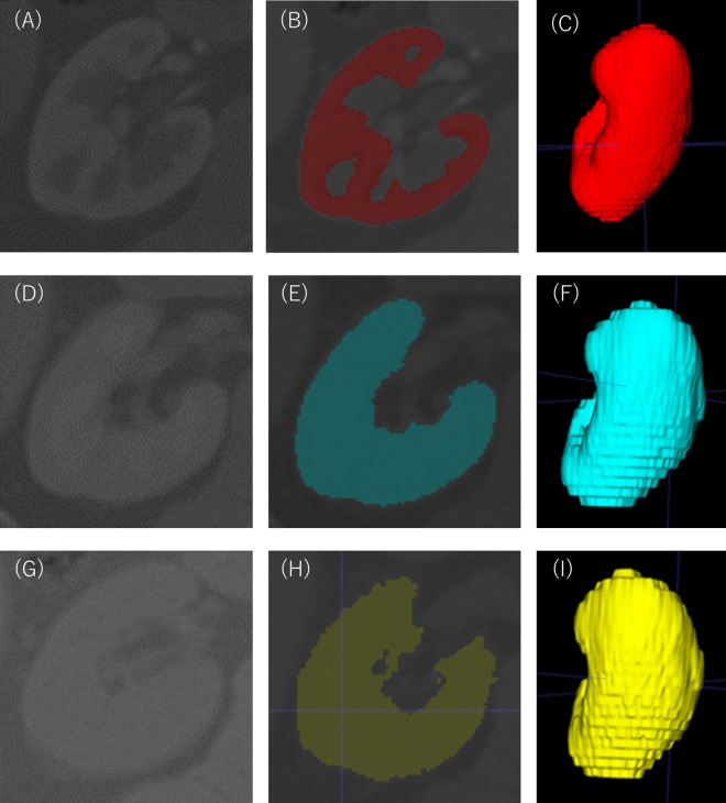
Figure 3.
Measurement of renal cortical volume and renal parenchymal volume. A three-dimensional cortical image was semi-automatically constructed from the contrast-enhanced images obtained in the arterial phase after contrast media infusion (A–C). A three-dimensional parenchymal image was semi-automatically constructed from the contrast-enhanced images obtained in the venous phase after contrast media infusion (D–F). As with the venous phase, a three-dimensional image was semi-automatically constructed from the image of specific density areas of kidney obtained from unenhanced CT images (G–I). Renal artery, renal vein, renal pelvis, ureter, renal sinus, fat in renal sinus, adjacent tissue and adjacent organs were excluded from all the created images.

