Figure 2.
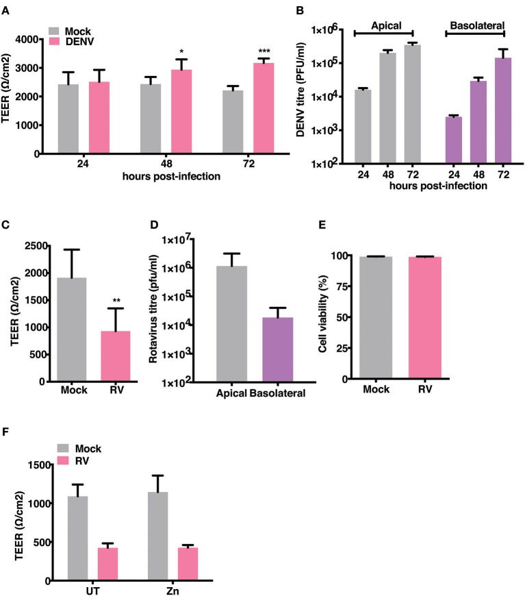
Effect of RV and DENV infection on epithelial barrier functions. Caco-2 cells were grown on transwell inserts for 4 days. Cells were infected with RV and DENV-2 at 0.5 and 10 MOI, respectively and TEER was measured. (A) TEER of DENV infected Caco-2 cells compared to mock measured till 72 h pi (B) Representative DENV titres determined by plaque assay and represented as pfu/ml at indicated time points. (C) TEER of RV infected Caco-2 cells compared to mock at 16 h pi (D) Representative RV titres determined by plaque assay and represented as pfu/ml at 16 h pi. (E) Viability of Caco-2 cells infected with RV as compared to mock measured by live dead staining using flow cytometry (F) TEER at 16 h pi of Caco-2 cells grown in transwells and infected with RV followed by addition of 50 μM ZnSO4. Data are from at least two independent experiments and represent mean ± SD. *p < 0.05, **p < 0.01, ***p < 0.001.

