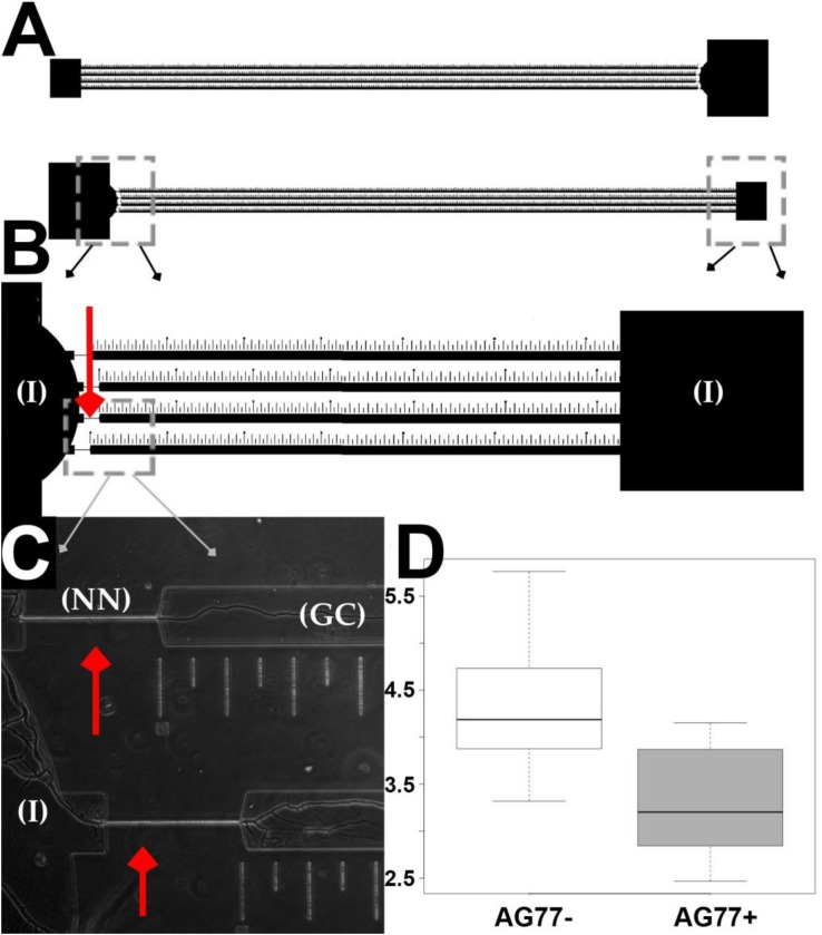
FIGURE 4.
Microfluidic device design and validation of fungal growth rate data. (A) Two chambers running parallel on a mounted slide. (B) Narrow necks indicated by red arrows running from inoculation chambers (I) to growth chamber etched with ruler in micrometers for single hyphal growth quantification. (C) M. elongata growing through narrow necks (NN) from inoculation chambers (I) into growth chamber (GC), imaged from device mounted onto a slide and filled with P20 liquid media. (D) Comparative microfluidic growth rate data (extrapolated to mm/day) for M. elongata with and without Mycoavidus cysteinexigens (AG77+ and AG77–).

