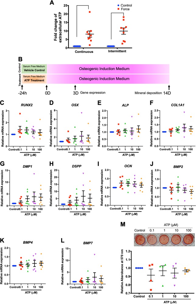
Fig. 3. ATP priming did not influence osteogenic differentiation in hPDLs.
Cells were treated with CCF or ICF in serum-free media for 24 h. Extracellular ATP was evaluated using an ATP assay in culture medium (a). Schematic diagram of the experimental plan of the ATP priming was illustrated (b). Cells were exposed to ATP for 24 h in serum-free medium. Thereafter, the culture medium was changed to osteogenic medium. Osteogenic marker gene expression was determined using real-time polymerase chain reaction at day 3 after osteogenic induction (c–l). Mineral deposition was examined using alizarin red s staining at day 14 (m). The normalized absorbance of the solubilized dye. Bars indicate a significant difference between conditions

