Figure 1.
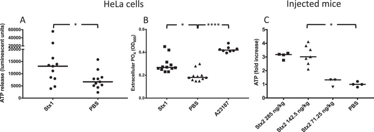
Shiga toxin induces release of ATP in vitro and in vivo. (A) HeLa cells (n = 11) were stimulated with Shiga toxin 1 (Stx1) or PBS and the ATP content was measured after 5 min. The median extracellular ATP content is depicted as the bars. (B) HeLa cells were stimulated with Stx1 (n = 12), A23187 calcium ionophore (n = 8) or PBS (n = 11). The supernatant was collected and free phosphate groups were measured. Data is depicted as median absorbance at OD600 (denoted by the bars), correlating to free phosphate groups in the supernatant. (C) Mice were injected with Stx2 at a concentration of 285 (n = 4), 142.5 (n = 7) or 71.25 ng/kg (n = 3) or with PBS as the control. Mice injected with Stx2 142.5 ng/kg had significantly higher plasma ATP levels compared to PBS control mice. A similar trend was seen for mice injected with Stx2 285 ng/kg, although the difference did not achieve statistical significance. Mice injected with Stx2 71.25 ng/kg had plasma ATP levels comparable to PBS control mice. The median is denoted by the bar. *P < 0.05, ****P < 0.0001, two-tailed Mann-Whitney test (panel A) and Kruskal-Wallis test (panels B and C).

