. 2019 Sep 25;54(3):236–240. doi: 10.5045/br.2019.54.3.236

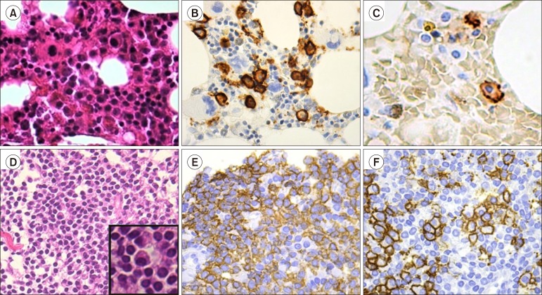
Fig. 2. (A–C) Neoplastic cells in the bone marrow in Case 1. (A) Hematoxylin and eosin staining: diffused invasion of the neoplastic cells was observed in the bone marrow biopsy specimen (original magnification ×400). (B) Staining with CD138: The cytoplasm of the neoplastic lymphoplasmacytes and plasmacytes was strongly positive for CD138 (×400). (C) Staining with CD20: The cytoplasm of the neoplastic small lymphocytes and lymphoplasmacytic cells were strongly positive for CD20 (×630). (D–F) The neoplastic cells of the mediastinum tumor mass in Case 2. (D) Hematoxylin and eosin staining: Neoplastic lymphocytes, lymphoplasmacytes, and plasmacytes were noted in the mediastinum tumor specimen (×400). (E) Staining with CD20: The cytoplasm of the neoplastic lymphocytes was strongly positive for CD20 (×400). (F) Staining with CD138: The cytoplasm of the neoplastic lymphoplasmacytes and plasmacytes was strongly positive for CD138 (×400).

Fig. 2