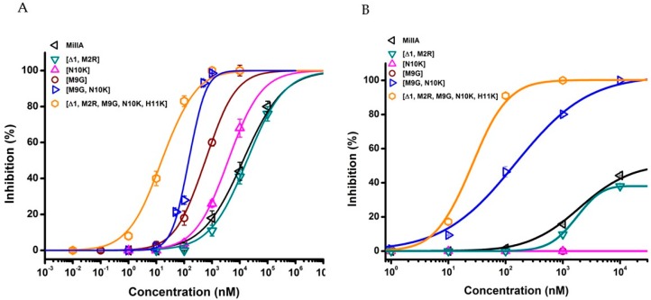
Figure 4.
Concentration curves on α1β1γδ (A) and α1β1δε (B) nAChR. The percentage of inhibition of MilIA, MilIA [M9G], MilIA [N10K], MilIA [∆1,M2R], MilIA [M9G, N10K], and MilIA [∆1,M2R, M9G, N10K, H11K] at α1β1γδ (left) and α1β1δε (right) nAChR was plotted against the logarithm of the different concentrations tested and fitted with the Hill equation. The visualized error bars represent the standard error of the mean (S.E.M). All of the experiments were repeated at least three times (n ≥ 3).

