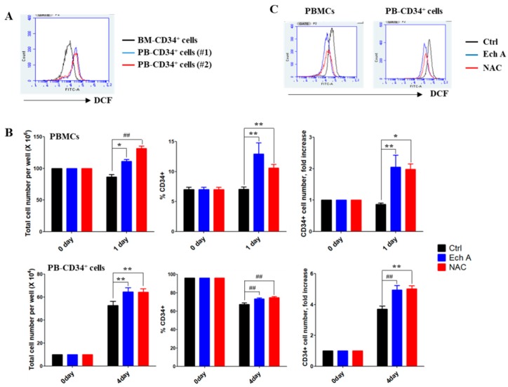
Figure 1.
Ech A increases PB-CD34+ cell expansion by inhibiting reactive oxygen species’ (ROS) generation. (A) Cells were treated with 10 μM 2′7′-dichlorodihydro-fluorescein diacetate for 30 min. The values in the 2′7′-dichlorofluorescein histograms indicate MFI (mean fluorescence intensity). (B,C) Cells were treated with 10 μM Ech A for 1 day (PB mononuclear cells, PBMCs) or 4 days (PB-CD34+ cells). For NAC treatment, cells were treated with 5 mM NAC for 4 h, washed, suspended in complete medium, and incubated for an additional 1 day (PBMCs) or 4 days (PB-CD34+ cells). (B) Total cell number was measured using the ADAM-MC automated mammalian cell counter (NanoEntech, Seoul, Korea). For flow cytometric immunophenotypic analysis, cells were stained with CD34-PE, CD38-FITC, CD45-APC, and 7-AAD. Each value was expressed as the mean ± SEM of three independent experiments. (C) Cells were treated with 10 μM 2′7′-dichlorodihydro-fluorescein diacetate for 30 min and ROS levels were subsequently measured usingthe flow cytometer.

