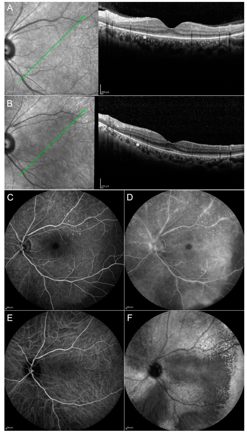
Figure 3.
Case 1, acute syphilitic posterior placoid chorioretinitis (ASPCC). Patients affected by ASPCC show typical outer retinal abnormalities on OCT with ellipsoid zone disruption (A) (asterisk), partially resolved after the treatment (B). FA reveals early hypofluorescence and late hyperfluorescence corresponding to the lesions (C,D). ICGA shows hypocyanescence until the late stages of the examination (E,F). Green arrow: plane and orientation of the optical coherence tomography (OCT) line scan.

