Figure 2.
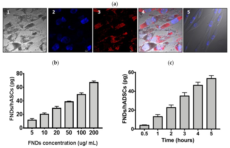
Characterization of fluorescent nanodiamond (FND)-labeled human adipose-derived stem/stromal cells (hASCs). (a) hASCs were incubated with 100 μg/mL for 4 h. The fluorescent images of FND-labeled hASCs were obtained by a fluorescent microscope. Cell nuclei were stained by 4′,6-diamidino-2-phenylindole (DAPI). The images were shown: (1) bright-field, (2) DAPI, (3) FNDs, (4) the merged image, and (5) hASCs without FNDs. Scale bar: 20 μm. (b) hASCs were incubated with FNDs at concentrations of 10–200 μg/mL for four hours. Quantification of FNDs in the cells was performed and analyzed by magnetically modulated fluorescence (MMF). (c) hASCs were incubated with FNDs in a time-course manner (half to five hours) at 100 μg/mL. The intracellular FNDs were subsequently analyzed by MMF.

