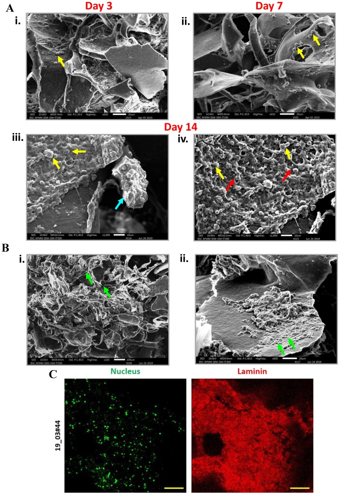
Figure 2.
Detection of cell clusters and extracellular matrix (ECM) deposition in 3D PCL scaffolds. (Ai–iv) SEM micrographs showed the presence of increasing numbers of cells in day 3, day 7 and day 14 cultures of red blood cell (RBC)-depleted nucleated cell pellet of patient samples cultured in 3D PCL scaffold (yellow arrows). Formation of clusters (iii) (blue arrow) and the presence of intercellular contacts (iv) (red arrows) was detected after 14 days of culture. Scale bar represents 20 μm. Images are representative of 6 patient samples. (B) Scanning electron microscopy analysis revealed the deposition of thread-like (i) and sheet-like (ii) ECM (green arrows). Images are representative of 4 patient samples. (C) Cells were immunostained for ECM protein Laminin (red); nucleus counterstained with Hoechst 33342 (pseudocoloured green). Imaging was performed using confocal microscope and maximum intensity projections are shown. Scale bar represents 100 μm. Minus primary antibody served as negative control and did not show staining; data not shown.

