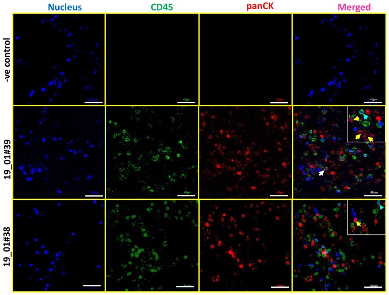
Figure 3.
Identification of CK-positive and CD45-negative CTCs in breast cancer patient blood samples cultured in 3D PCL scaffold. Immunostaining shows the presence of panCK+/CD45− CTCs in day 14 cultures of patient blood samples cultured in 3D PCL scaffolds. Top panel shows negative control (minus primary antibody). Middle and bottom panels show immunostaining done on two different patient samples. Yellow arrows indicate panCK+ and CD45 negative CTCs while blue arrows indicate CD45 positive leucocyte lineage cells. White arrow shows CK+ cells surrounded by CD45+ cells. Imaging was performed using confocal microscope, and maximum intensity projections are shown. Inset shows higher magnification. Scale bar represents 40 μm. Images are representative of 7 independent patient samples.

