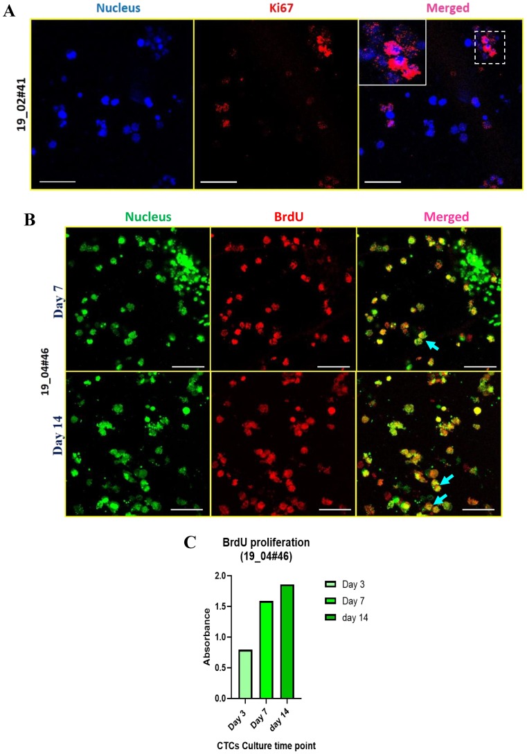
Figure 4.
Active cell proliferation in breast cancer patient-derived cells cultured in 3D PCL scaffolds. (A) Fluorescence images of Ki-67 positive patient blood cells harvested after 14 days of culture in 3D PCL scaffolds. Cells were stained for nucleus (Hoechst 33342, blue), Ki-67 (red). Images are representative of 2 independent patient samples. (B) Patient-derived cells were exposed to a single pulse of 50 μM BrdU on day 7 and day 14 of culture and harvested after 48 h for immunostaining for BrdU incorporation. Fluorescence images show BrdU-positive cells (red), stained for nucleus (Sytox green); blue arrows show the merge. Imaging was performed using confocal microscope and maximum intensity projections are shown. Scale bar represents 40 μm (A,B). (C) Graph shows BrdU incorporation in patient-derived cells over time (day 3, day 7, day 14) in culture as measured by colorimetry. Images are representative of 2 independent patient samples.

