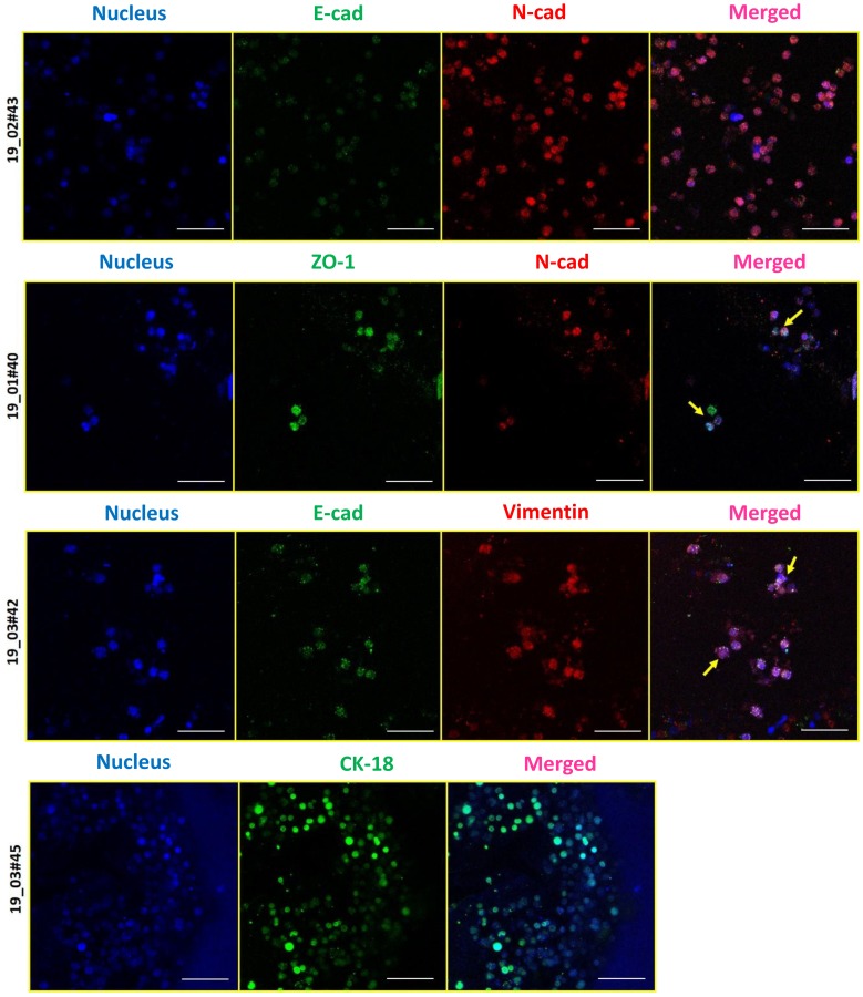
Figure 5.
E-M characterization of CTCs derived from blood samples of breast cancer patients. Immunostaining revealed the presence of epithelial (E)-type CTCs expressing E-cad, ZO-1, or CK18 (green), and mesenchymal (M)-type CTCs expressing vimentin or N-cad (red). Cells were stained for nucleus (Hoechst 33342, blue). Imaging was performed using confocal microscope and maximum intensity projections are shown. Scale bar represents 40 μm. Images are representative of 4 independent patient samples.

