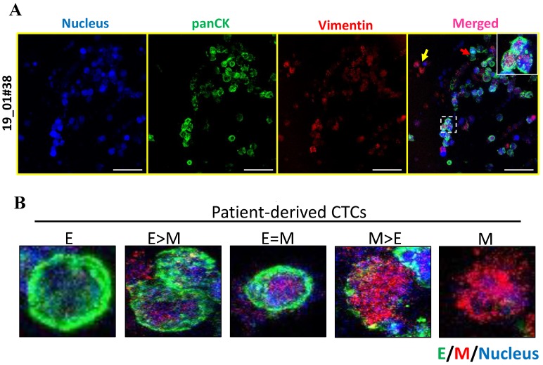
Figure 6.
E-M heterogeneity and intermediate phenotypes in breast cancer patient-derived CTCs. (A) Co-staining for panCK and vimentin in patient-derived CTCs revealed the presence of marked heterogeneity with respect to E and M marker expression. (B) Representative fluorescence images of 5 different types of CTCs: E (exclusively), E > M, E = M, M > E and M (exclusively). Cells were counterstained for nucleus (Hoechst 33342, blue), E-type markers (green), and M-type markers (red). Imaging was performed using confocal microscope and maximum intensity projections are shown. Scale bar represents 40 μm (A). Images are representative of 2 independent patient samples.

