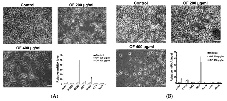
Figure 3.
Differentiation induction of human MG cells after treatment with oligo-fucodian (OF). (A) GBM8401 and (B) U87MG cells were treated with OF for seven days. Morphology of the MG cells was examined by inverted phase contrast microscopy. Scale bar is 50 μm. Expression of differentiation marker genes was analyzed by quantitative PCR. Astrocyte markers: GFAP and S100B; oligodentrocyte markers: Olig2 and MBP; neuron markers: MAP2, TUJ1 and NeuN. Data were expressed as mean ± standard error.

