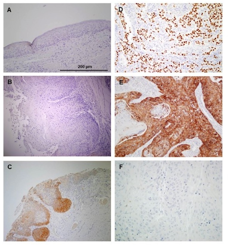
Figure 1.
Immunohistochemical analysis of NANOG expression in oral epithelial dysplasias and oral squamous cell carcinoma (OSCC). The normal adjacent epithelium exhibited negative staining (A). Representative examples of oral dysplasias showing negative (B), and positive NANOG staining (C), human seminoma as a positive control (D). Examples of oral squamous cell carcinomas with positive (E), and negative NANOG staining (F). Magnification 200×. Scale bar 200 µm.

