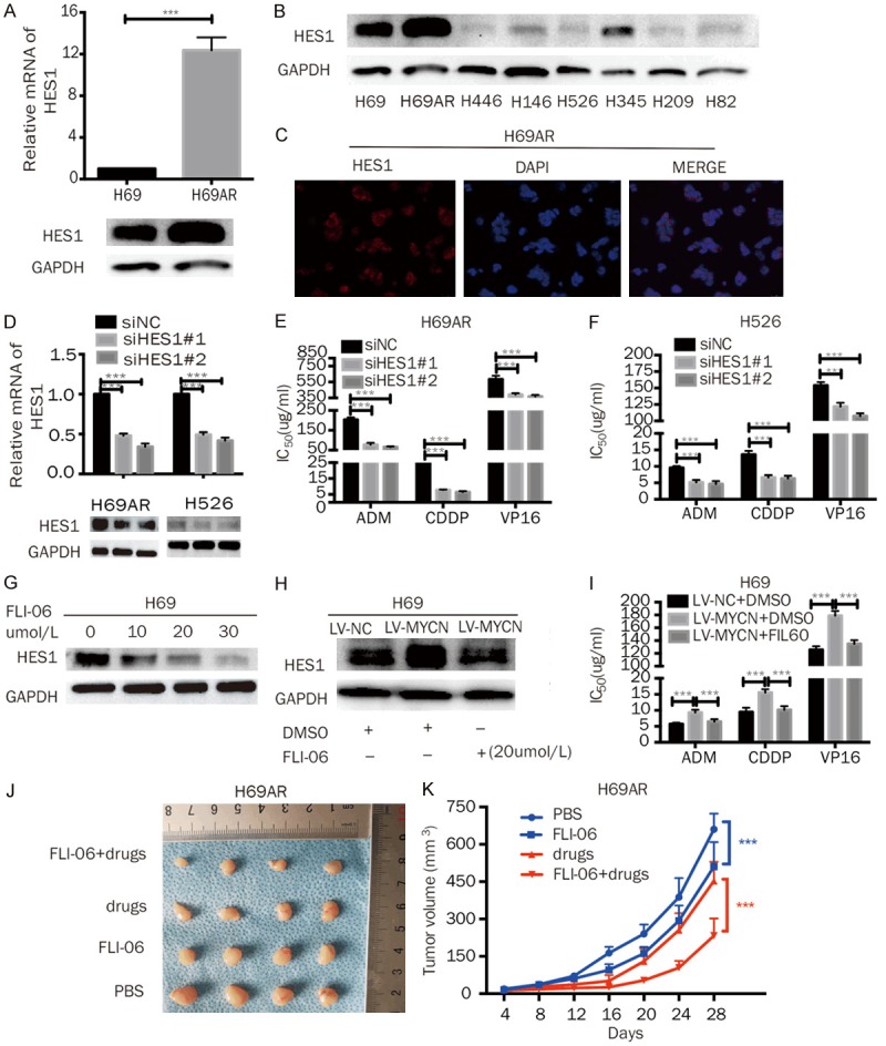
Figure 5.

MYCN promotes the chemoresistance of SCLC through HES1. A. RT-qPCR and Western blot analysis of HES1 expression in H69 and H69AR cells. B. Western blot analysis of HES1 expression in eight SCLC cell lines (H69, H69AR, H446, H146, H526, H345, H209, and H82). C. The cellular localization of HES1 was confirmed by immunofluorescence staining of H69AR cells. D. RT-qPCR and Western blot analysis of MYCN in H69AR and H526 cells transfected with siRNA against HES1. E, F. CCK-8 assays showed that HES1 knockdown decreased the IC50 values of chemotherapeutic agents (ADM, CDDP, and VP-16) in H69AR and H526 cells. G. Western blotting analysis revealed that 24 h of FLI-06 treatment inhibited HES1 expression in a dose-dependent manner. H. Western blotting showed that MYCN overexpression increased HES1 expression; however, the increased HES1 expression was diminished by FLI-06. I. A CCK-8 assay showed that the IC50 values were significantly increased in H69-LV-MYCN cells compared with those in the control cells, and the inhibition of HES1 by FLI-06 in MYCN-overexpressing cells could abate the increase in the IC50 values mediated by MYCN upregulation. J. Effect of FLI-06 with or without chemotherapy drugs (CDDP+VP16) on subcutaneous tumor growth injected with H69AR cells (n = 4 per group). K. Growth curve of tumor volumes of H69AR cells using FLI-06 with or without drugs (CDDP+VP16). Error bars indicate the mean ± SD from three independent experiments; ***P < 0.05.
