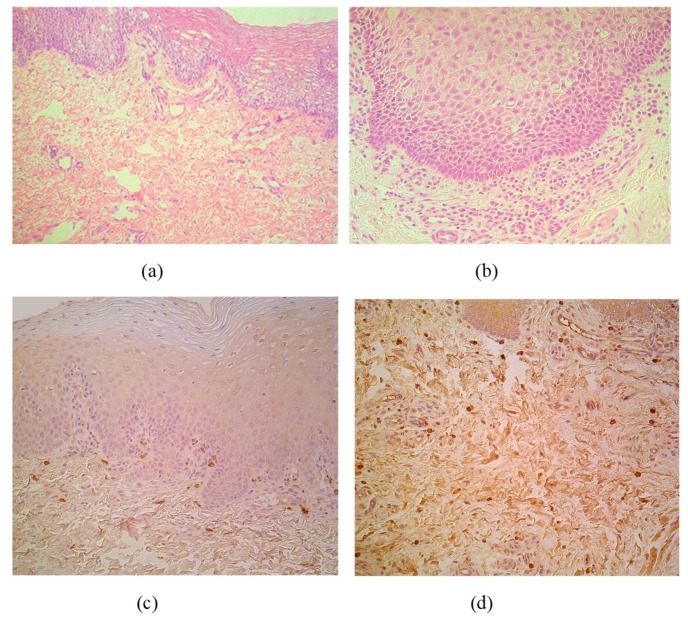
Figure 1.
(a–d) Micrographs of cleft lip affected lip in children. (a) Note patchy vacuolization of the lip epithelium (keratosis seborrhoea) in a 3-month-old child. Hematoxylin and eosin, X 200; (b) Visible proliferation of the basal epitheliocytes and subepithelial inflammation in a 3-month-old child. Hematoxylin and eosin, X 250. (c) MIP-1ß immunoreactive macrophages are located strictly beneath the basal membrane of 5-month-old child. MIP-1ß IMH, X 200; (d) Note diffuse distribution of MIP-1ß immunoreactive macrophages in subepithelial connective tissue of 4-month-old child. MIP-1ß IMH, X 200.

