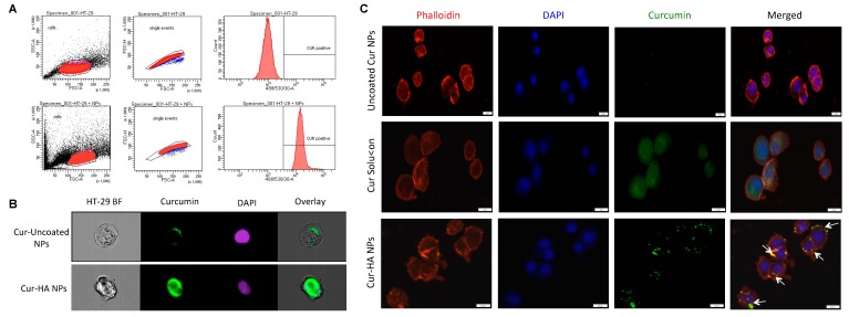
Figure 5.
Hyaluronan functionalization increases the nanoparticles interaction, uptake in HT-29 cells. Flow cytometry histogram shows alone HT-29 cells, HT-29 cells treated with Cur-HA-NPs signal after 3 h incubation (A). Representative ImageStream flow cytometry images show HA functionalized particles has effective uptake in HA-29 cells (B). Representative slide scanner images of uncoated Cur NPs, Cur solution and Cur-HA NPs after 3 h incubation, HA functionalized NPs showing more cellular interactions and uptake compared to uncoated NPs; nuclei (blue) stained with DAPI, cytoskeleton (red) stained with rhodamine-phalloidin, curcumin (green) (C).

