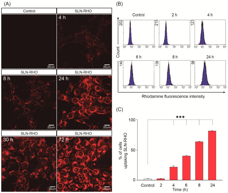
Figure 2.
Uptake of SLN–RHO into HEI-OC1 cells. (A) HEI-OC1 cells were incubated with 250 µg/mL of rhodamine-loaded SLN (SLN–RHO). Fluorescence intensity of incorporated SLN–RHO was detected by confocal microscopy at the times indicated. Representative microphotographs from three independent experiments are shown. (B) HEI-OC1 cells were incubated with SLN–RHO (250 µg/mL) for different times, cells were then collected, and the fluorescent intensity of incorporated SLN–RHO was detected by flow cytometry. The plots shown are representative of three independent experiments, whose quantification is shown in (C). Data are shown as the mean ± SEM from three independent experiments. One-way ANOVA was used to determine statistical significance; ***p < 0.001.

