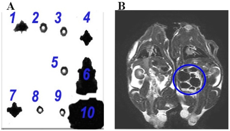
Figure 5.
A: The T2-weighted MRI of in vitro samples. 1: DPSCs labeled with 3.5 mg/mL of SPIONs showing a hypointense signal of the iron oxide contrast, 2: Non-labeled DPSCs without any hypointense signal for the iron oxide contrast, 3: 15% agarose gel lacking the hypointense signal of the iron oxide contrast, 4: 5 mg/mL of SPIONs in 15% gel revealing the hypointense signal of the iron oxide contrast, 5: 15% gel without any hypointense signal of the iron oxide contrast, 6: 5 mg/mL SPIONs demonstrating the hypointense signal of the iron oxide contrast, 7: DPSCs labeled with 3.5 mg/mL SPIONs denoting the hypointense signal of the iron oxide contrast, 8: DPSCs labeled with 0.35 mg/mL SPIONs in absence of any hypointense signal of the iron oxide contrast, 9: H2O lacking the hypointense signal of the iron oxide contrast, and 10: Non-coated iron oxide particle exhibiting an extremely hypointense signal of the iron oxide contrast. (B) The T2-weighted images of DPSCs labeled with 3.5 mg/mL SPIONs injected intraperitoneally with hypointense signal of the iron oxide contrast, in the right picture, compared to non-labeled DPSCs, in the left picture, lacking the hypointense signal (rats in supine position).

